
Chest pain, a strong family history of heart disease, or an uncertain risk picture can leave you wondering what’s really going on inside your heart. For many patients, the answer increasingly comes from cardiac CT, a detailed, non-invasive scan that lets a cardiologist look directly at the heart’s arteries. For people across Brisbane’s bayside and eastern suburbs, BK Heart in Alexandra Hills uses cardiac CT as an integral part of everyday cardiac assessment, particularly when investigating chest pain.
This guide explains what cardiac CT imaging is, why it might be recommended, what the process involves, and how the results help shape your care.
Cardiac CT (computed tomography) is an advanced imaging technique that uses X-rays and computer processing to create detailed, cross-sectional pictures of the heart and its blood vessels. Two related types are commonly used:
Unlike an invasive angiogram, cardiac CT doesn’t require a catheter to be threaded through your arteries. It’s a non-invasive test, which is one reason it has become such a valuable tool in modern cardiology. The Heart Foundation provides accessible background on the range of heart tests used in Australia, including imaging.
Your cardiologist may recommend cardiac CT for several reasons. It’s especially valuable in the assessment of chest pain, where it can help determine whether the coronary arteries are healthy or whether disease is present. Other common reasons include:
At BK Heart, cardiac CT is an integral part of day-to-day cardiac services, and it’s particularly useful in the assessment of chest pain. You can see how it fits within the clinic’s broader imaging and diagnostic services, accessible to patients from Wynnum, Manly, Tingalpa, Cannon Hill, Capalaba and Cleveland.
A common question is where and how the scan is performed. At BK Heart, the cardiac CT scans themselves are performed off site at an imaging facility, and the images are then carefully reviewed by an experienced cardiologist to ensure accurate interpretation and clear clinical direction.
This two-step approach has a clear benefit: the scan is acquired using dedicated CT equipment, while the interpretation, the part that translates images into meaning for your health, is handled by a cardiologist who understands your full clinical picture.
Here’s what the experience typically involves:
Before the scan. You may be given specific instructions to prepare, which can include avoiding caffeine for a period beforehand, as caffeine can affect your heart rate. In some cases, medication is given to gently slow the heart rate during the scan, because a steady, slower rhythm produces clearer images. Your care team will explain any preparation that applies to you.
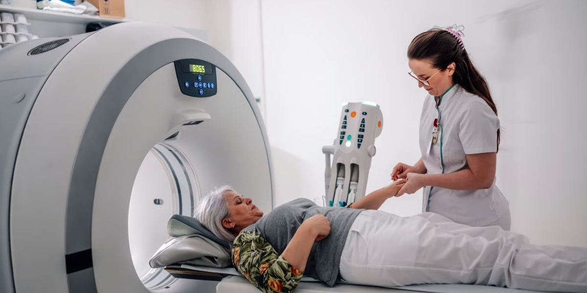
During the scan. You lie on a table that moves through the CT scanner, which is shaped like a large ring rather than an enclosed tunnel. Small electrodes monitor your heartbeat so the images are timed precisely to your heart’s motion. If contrast dye is used, it’s given through a small cannula in your arm and may cause a brief warm sensation. The scan itself is quick, and you’ll be asked to hold your breath for a few seconds at the key moment.
After the scan. The imaging is sent to your cardiologist for review.
Once your scan has been assessed, the results are discussed with you in a dedicated consultation. At BK Heart, this is an important part of the process the team takes the time to explain what the findings mean, answer your questions, and make sure you feel informed and supported.
Cardiac CT is reviewed by Dr Neelabh Sharma, a consultant cardiologist (MBBS, FRACP) with a special interest in multimodality cardiac imaging, including cardiac CT. From there, your care is tailored to your individual needs, risk factors, and symptoms. Depending on the findings, management decisions may include adjusting your medications, organising additional functional testing such as a stress echocardiogram when appropriate, or considering referral for further procedures if required.
The goal isn’t just to produce an image, but to translate that image into a clear plan for protecting your heart.
Cardiac CT does involve a small dose of X-ray radiation, and where contrast dye is used there are some considerations for people with kidney concerns or dye allergies. These factors are part of why the test is ordered by a specialist who weighs the benefits against any risks for your individual situation. Modern cardiac CT techniques are designed to keep radiation doses as low as reasonably achievable while still producing diagnostic-quality images. Your cardiologist will discuss whether cardiac CT is appropriate and suitable for you.
If you’ve been experiencing chest pain, have an uncertain cardiovascular risk picture, or simply want a thorough specialist assessment of your heart health, cardiac CT can provide valuable answers. BK Heart in Alexandra Hills offers cardiac CT as part of comprehensive cardiac care for patients across Brisbane’s bayside and eastern suburbs including Lota, Manly West, Wynnum West, Hemmant, Murarrie, Wakerley, Gumdale and Thornlands with specialist interpretation and a dedicated consultation to explain your results.
Speak with your GP about a referral, then contact our team to find out more.
BK Heart — The Specialist Cardiac Centre
Shop 38/71 Cambridge Drive, Alexandra Hills, QLD 4161
Phone: (07) 3733 1536 · Email: info@bkheart.com.au
Monday to Friday, 8:30 AM – 4:30 PM